What Occurs to a Neuron When It Receives a Chemical Message
Learning Objectives
- Describe the structure and function of neurons
- Interpret an activity potential graph and explain the molecular mechanisms underlying each footstep of the activity potential
- Describe the structure and function of neuronal synapses and the role of neurotransmitters at the synapse
Neurons and Glial Cells
The information below was adapted from OpenStax Biology 35.one and Khan Academy AP Biology The neuron and nervous system. All Khan Academy content is available for gratis at www.khanacademy.org
The nervous arrangement is made upwards of neurons, the specialized cells that tin can receive and transmit chemical or electrical signals, and glia, the cells that provide support functions for the neurons. A neuron can be compared to an electric wire: it transmits a signal from one place to another. Glia can exist compared to the workers at the electrical company who brand sure wires go to the right places, maintain the wires, and accept down wires that are broken. Recent prove suggests that glia may also aid in some of the signaling functions of neurons.
Neurons communicate via bothelectrical signals andchemic signals. The electrical signals areaction potentials, which transmit the data from one of a neuron to the other; the chemic signals areneurotransmitters, which transmit the information from ane neuron to the next. An action potential is a rapid, temporary change in membrane potential (electrical accuse), and information technology is acquired bysodium rushing to a neuron andpotassium rushing out. Neurotransmitters are chemical messengers which are released from 1 neuron as a result of an action potential; they cause a rapid, temporary modify in the membrane potential of the adjacent neuron to initiate an action potential in that neuron.
Parts of a Neuron
Similar other cells, each neuron has a jail cell body (or soma) that contains a nucleus and other cellular components. Neurons likewise contain unique structures, dendrites and axons, for receiving and sending the electrical signals that make neuronal communication possible:
- Dendrites:are tree-similar structures that extend away from the cell torso to receive neurotransmitters from other neurons. Some types of neurons do not accept any dendrites, some types of neurons have multiple dendrites. Dendrites can have pocket-sized protrusions called dendritic spines, which further increase surface area for possible connections with other neurons.
- Synapses:Dendrites receive signals from other neurons at specialized junctions called synapses. There is a small gap betwixt two synapsed neurons, where neurotransmitters are released from 1 neuron to pass the point to the next neuron.
- Axon hillock:Once a point is received past the dendrite, it and then travels to the jail cell body. The prison cell body contains a specialized structure, the axon hillock that "integrates" signals from multiple synapses and serves as a junction between the cell body and an axon.
- Axon:An axon is a tube-like construction that propagates the integrated signal to specialized endings chosen axon terminals. The axon carries the action potential to the next neuron. Neurons usually take one or two axons. Some axons are covered with myelin, which acts as an insulator to minimize dissipation of the electrical point as it travels down the axon, greatly increasing the speed on conduction. This insulation is important as the axon from a human being motor neuron can exist as long as a meter, from the base of the spine to the toes. The myelin sheath is non actually function of the neuron, and is produced by glial cells. Along the axon at that place are periodic gaps in the myelin sheath chosen nodes of Ranvier, whichare sites where the signal is "re-charged" equally information technology travels along the axon.
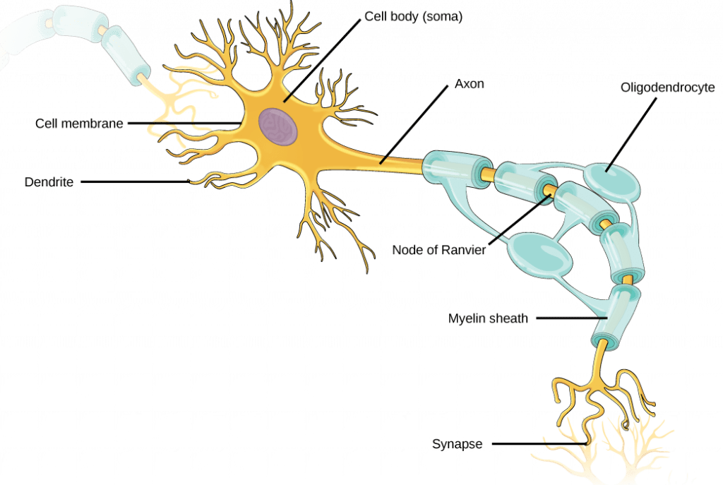
Neurons incorporate organelles common to many other cells, such as a nucleus and mitochondria. They also accept more specialized structures, including dendrites and axons. Paradigm credit: OpenStax Biological science
It is important to annotation that a single neuron does not act alone: neuronal communication depends on the connections that neurons make with i another (too as with other cells, similar musculus cells). Dendrites from a single neuron may receive synaptic contact from many other neurons. For example, dendrites from a neurons in the cerebellum of the brain are thought to receive contact from as many as 200,000 other neurons.
Glia
While glia are ofttimes idea of every bit the supporting cast of the nervous system, the number of glial cells in the brain actually outnumbers the number of neurons by a factor of ten. Neurons would exist unable to function without the vital roles that are fulfilled past these glial cells. Glia guide developing neurons to their destinations, buffer ions and chemicals that would otherwise harm neurons, provide myelin sheaths effectually axons, and modulate advice between nervus cells. When glia do non function properly, the result can exist disastrous; most encephalon tumors are caused by mutations in glia.
There are several dissimilar types of glia with different functions. They include:
- Astrocytes: provide nutrients and other substances to neurons, regulate the concentrations of ions and chemicals in the extracellular fluid, and provide structural support for synapses; also form the blood-brain bulwark, which blocks archway of toxic substances into the brain.
- Satellite glia:nutrients and structural support for neurons in the peripheral nervous system (PNS).
- Microglia: allowed cells of the primal nervous system (CNS); scavenge and dethrone dead cells and protect the brain from invading microorganisms.
- Oligodendrocytes:course myelin sheaths around axons in the CNS; one axon can be myelinated by several oligodendrocytes, and 1 oligodendrocyte tin can provide myelin for multiple neurons.
- Schwann cells: class myelin sheaths around axons in the PNS; dissimilar oligodendrocytes, a single Schwann cell provides myelin for simply one axon equally the unabridged Schwann cell surrounds the axon.
- Ependymal cells: line fluid-filled ventricles of the brain and the cardinal culvert of the spinal cord; aid circulate cerebrospinal fluid, which serves as a absorber for the brain.

Glial cells support neurons and maintain their environment. Glial cells of the (a) cardinal nervous system include oligodendrocytes, astrocytes, ependymal cells, and microglial cells. Oligodendrocytes class the myelin sheath around axons. Astrocytes provide nutrients to neurons, maintain their extracellular environment, and provide structural back up. Microglia scavenge pathogens and expressionless cells. Ependymal cells produce cerebrospinal fluid that cushions the neurons. Glial cells of the (b) peripheral nervous system include Schwann cells, which form the myelin sheath, and satellite cells, which provide nutrients and structural back up to neurons. Epitome credit: OpenStax Biology
Communication Between Neurons
The data below was adapted from OpenStax Biological science 35.2 and OpenStax Anatomy & Physiology iii.1
All functions performed by the nervous system – from a simple motor reflex to more avant-garde functions like making a memory or a determination – require neurons to communicate with one another. While humans apply words and body language to communicate, neurons use electrical and chemical signals. Just like a person in a committee, ane neuron usually receives and synthesizes letters from multiple other neurons before "making the conclusion" to transport the message on to other neurons.
Neurons communicate via both electrical and chemic signals. A neuron receives input from other neurons and, if this input is potent enough, the neuron volition send the signal to downstream neurons. Transmission of a bespeak between neurons is mostly carried past a chemical called a neurotransmitter. Transmission of a bespeak inside a neuron (from dendrite to axon concluding) is carried by a brief reversal of the resting membrane potential called an action potential. This advice is possible because each neuron has a charged cellular membrane (a voltage difference betwixt the within and the exterior), and the charge of this membrane tin can change in response to neurotransmitter molecules released from other neurons and environmental stimuli. The 3 full general phenomena required for communication between neurons are:
- Resting potential: the membrane potential (electric charge) in a neuron that isnon currently transmitting a signal
- Action potential: a brief depolarizaiton (reduction in magnitude of the charge) along the neuron's axon; action potentials are all-or-nothing (they do not have degrees of magnitude)
- Neurotransmitters: the chemic messengers that communicate between side by side neurons; release of neurotransmitters from one neuron will either help depolarize or hyperpolarize (increase the magnitude of the charge) the adjacent neuron, making an action potential either more or less likely to occur in the adjacent neuron
We'll hash out each of these three components in plow.
i. The Resting Potential
The lipid bilayer membrane that surrounds a neuron is impermeable to charged molecules or ions. To enter or exit the neuron, ions must pass through special proteins called ion channels that bridge the membrane and regulate the relative concentrations of different ions inside and exterior the cell. Cells can employ energy to preferentially movement certain ions either inside or exterior of the membrane, setting up a deviation in ion accuse across the membrane, where one side is relatively more than negative and the other side is relatively more positive. The deviation in full charge between the within and exterior of the cell is called the membrane potential.
The membrane potential of a neuron at residue is negatively charged: the within of a cell is approximately 70 millivolts more negative than the exterior (-lxx mV, notation that this number varies by neuron type and by species). This voltage is chosen the resting membrane potential; information technology is caused by differences in the concentrations of ions inside and outside the prison cell. The resting potential is established and maintained by two main processes: an ATP-powered ion aqueduct called thesodium-potassium pump, and a passive ion aqueduct called thepotassium leak channel.
The sodium-potassium pump, which is too called Na+/K+ ATPase, transports sodium out of a cell while moving potassium into the cell. The Na+/K+ pump is an important ion pump found in the membranes of many types of cells. These pumps are particularly abundant in nerve cells, which are constantly pumping out sodium ions and pulling in potassium ions to maintain an electrical gradient across their jail cell membranes. An electrical slope is a difference in electrical charge across a space. In the example of nervus cells, for example, the electrical gradient exists between the inside and exterior of the cell, with the inside being negatively-charged (at around -70 mV) relative to the exterior. The negative electric slope is maintained because each Na+/Grand+ pump moves three Na+ ions out of the prison cell and two Grand+ ions into the cell for each ATP molecule that is used. This procedure is so important for nervus cells that it accounts for the majority of their ATP usage.
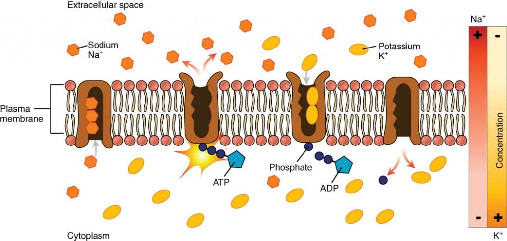
Powered by ATP, the sodium-potassium pump moves sodium and potassium ions in opposite directions, each against its concentration gradient. In a single cycle of the pump, iii sodium ions are extruded from and two potassium ions are imported into the cell. Image credit: OpenStax Beefcake & Physiology.
In addition to the sodium potassium pump, neurons possess potassium leak channels and sodium leak channels that allow the two cations to diffuse down their concentration gradient. However, the neurons have far more potassium leakage channels than sodium leakage channels. Therefore, potassium diffuses out of the cell at a much faster rate than sodium leaks in. Because more cations are leaving the cell than are entering, this causes the interior of the cell to exist negatively charged relative to the outside of the prison cell. Thus the combined effects of the sodium-potassium pump and the potassium leak channels is that the interior of the jail cell is more negative than the outside of the cell. It should also be noted that chloride ions (Cl–) tend to accrue outside of the cell because they are repelled by negatively-charged proteins within the cytoplasm.
| Ion Concentration Inside and Outside Neurons | |||
|---|---|---|---|
| Ion | Extracellular concentration (mM) | Intracellular concentration (mM) | Ratio outside/inside |
| Na+ | 145 | 12 | 12 |
| K+ | four | 155 | 0.026 |
| Cl– | 120 | iv | thirty |
| Organic anions (A-) | — | 100 | |
This video describes the part of the sodium/potassium pump and potassium leak channels in establishing and maintaining the membrane resting potential:
2. The Action Potential
When nosotros talk nigh neurons "firing" or existence "active," we're talking well-nigh the activeness potential:a brief, positive change in the membrane potential along a neuron's axon. When an action potential occurs, the neuron sends the point to the next neuron in the advice chain, and, if an action potential besides occurs in the side by side neuron, then the signal will continue being transmitted. What causes an activity potential? When a neuron receives a signal from some other neuron (in the grade of neurotransmitters, for most neurons), the signal causes a change in the membrane potential on the receiving neuron. The bespeak causes opening or closing ofvoltage-gated ion channels, channels that open up or close in response to changes in the membrane voltage. The opening of voltage-gated ion channels causes the membrane to undergo either ahyperpolarization, where the membrane potential increases in magnitude (becomes more negative) or adepolarization, where the membrane potential decreases in magnitude (becomes more positive). Whether the membrane undergoes a hyperpolarization or a depolarization depends on the type of voltage-gated ion channel that opened.
Not all depolarizations effect in an action potential. The point must cause a depolarization that is large plenty in magnitude to overcome thethreshold potential, or the specific voltage that the membrane must reach for an an activeness potential to occur. The threshold potential is usually about -55 mV, compared to the resting potential of near -70 mV. If the threshold potential is reached, then an activeness potential is initiated at theaxon hillock in the post-obit stages:
- Depolarization: voltage-gated sodium channels open quickly afterwards depolarization past the threshold potential. Equally sodium rushes into the axon (influx), the inside becomes relatively electrically positive (approximately +30 mV, compared to the initial resting potential of apprximately -70 mV).
- Repolarization: soon after the initial depolarization, the voltage-gated sodium channels close and remain airtight (and cannot exist opened) for about ane-2 milliseconds. Voltage-gated potassium channels then open, assuasive potassium to rush out of the axon (efflux), causing the membrane to repolarize (become more negative).
- Hyperpolarizaton: potassium continues leaving the axon to the signal that the membrane potential dips beneath the normal resting potential. Sodium channels return to their resting state, meaning they are fix to open again if the membrane potential again exceeds the threshold potential.
- Reset resting potential: The sodium-potassium pump and potassium leak channels reset the locations of sodium and potassium ions, reestablishing the membrane potential to allow some other action potential to burn down.
These steps are illustrated here:
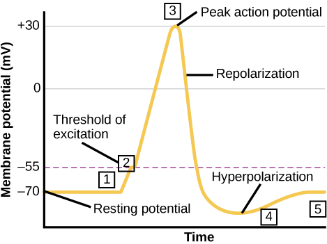
The formation of an action potential can be divided into 5 steps: (ane) A stimulus from a sensory cell or another neuron causes the target cell to depolarize toward the threshold potential. (2) If the threshold of excitation is reached, all Na+ channels open and the membrane depolarizes. (3) At the pinnacle activity potential, K+ channels open and Chiliad+ begins to get out the cell. At the same time, Na+ channels close. (4) The membrane becomes hyperpolarized equally K+ ions keep to exit the cell. The hyperpolarized membrane is in a refractory period and cannot fire. (5) The One thousand+ channels close and the Na+/Grand+ transporter restores the resting potential. Paradigm credit: OpenStax Biological science.
There are a few of import universal features of action potentials:
- The action potential travels down the axon, proceeding equally a wave of depolarization. The image above shows a trace of an action potential at a single indicate in the membrane of an axon; the same blueprint repeats down the entire length of the axon until it reaches the synapse, shown here:
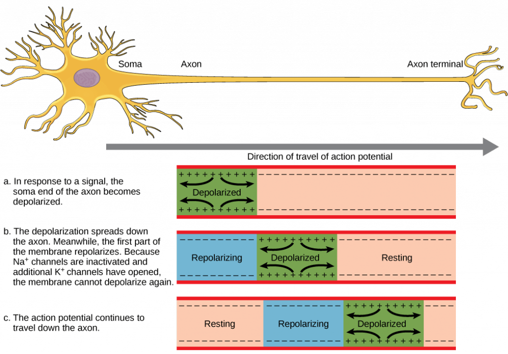
The activity potential is conducted down the axon as the axon membrane depolarizes, then repolarizes. Prototype credit: Openstax Biology.
- Action potentials always proceed in ane direction only, from the cell trunk (soma) to the synapse(south) at the end of the axon. Activity potentialsnever go backward, due to therefractory period of the voltage-gated ion channels, where the channels cannot re-open for a menses of 1-2 milliseconds after they have closed. The refractory period forces the action potential to travel only in 1 management.
- Action potentials exercise not vary inmagnitude or speed; they are"all-or-nothing."When a given neuron fires, the action potential e'er depolarizes to the same magnitude and e'er travels at the same speed along the axon. At that place is no such thing every bit a bigger or faster action potential. The parameter that tin can vary is the frequency of action potentials, or how many activity potentials occur in a given corporeality of time.
The video below provides a discussion of voltage-gated ion channels:
Here is a more detailed discussion of the action potential trace:
And an overview of action potential propagation:
Equally noted above, the magnitude or speed of the action potential for a given neuron never varies; even so, some neurons have faster activity potentials than others. In invertebrates, this departure is often due to axon diameter, where larger axons accept faster conduction of action potentials. In vertebrates, this difference is typically due to myelination of the neuron's axon. Myelin acts every bit an insulator that prevents electric current from leaving the axon; this increases the speed of action potential conduction. The nodes of Ranvier, illustrated beneath, are gaps in the myelin sheath forth the axon. These unmyelinated spaces are about one micrometer long and comprise voltage gated Na+ and Thousand+ channels. Catamenia of ions through these channels, particularly the Na+ channels, regenerates the activeness potential over and over again along the axon. This "jumping" of the action potential from one node to the side by side is called saltatory conduction . Nodes of Ranvier besides relieve free energy for the neuron since the channels only need to exist present at the nodes and not along the entire axon.
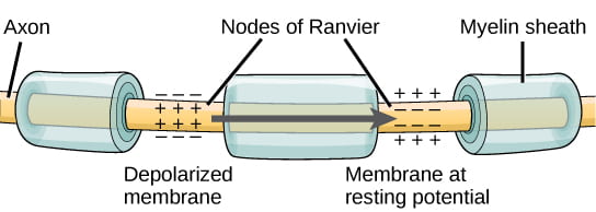
Nodes of Ranvier are gaps in myelin coverage along axons. Nodes comprise voltage-gated K+ and Na+ channels. Action potentials travel downwardly the axon by jumping from one node to the next. Image credit: OpenStax Biology
3. The Chemic Synapse and Neurotransmitters
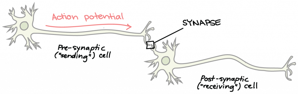
Neurons are non in direct physical contact with each other, only instead come into very close proximity at a structure chosen the synapse. The neuron sending a signal to the side by side is called thepresynaptic neuron, and the neuronreceiving a signal is called thepostsynaptic neuron, shown here:
Chemic transmission involves release of chemical messengers known as neurotransmitters. Neurotransmitters carry data from the pre-synaptic (sending) neuron to the mail service-synaptic (receiving) prison cell. Image credit: Khan University https://www.khanacademy.org/scientific discipline/biology/ap-biology/man-biology/neuron-nervous-system/a/the-synapse
There is a small gap between the two neurons called thesynaptic cleft, whereneurotransmitters are released by the presynaptic neuron to transmit the signal to the postsynaptic neuron, shown here:
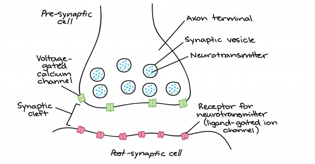
Inside the axon last of a sending cell are many synaptic vesicles. These are membrane-bound spheres filled with neurotransmitter molecules. There is a pocket-sized gap betwixt the axon terminal of the presynaptic neuron and the membrane of the postsynaptic cell, and this gap is chosen the synaptic crevice. Epitome credit: Khan Academy https://world wide web.khanacademy.org/science/biology/ap-biology/human-biology/neuron-nervous-arrangement/a/the-synapse
How does synaptic transmission work? In one case the activity potential reaches the end of the axon, it propagates into the pre-synaptic last where the following events occur in sequence:
- The action potential depolarizes the membrane and opens voltage-gated Na+ channels. Na+ ions enter the prison cell, farther depolarizing the presynaptic membrane.
- This depolarization causes voltage-gated Caii+ (calcium) channels to open in the presynaptic neuron, allowing calcium ions to enter the presynaptic neuron at the synpase.
- Calcium ions entering the presynaptic neuron jail cell initiate a signaling cascade that causes small membrane-leap vesicles, called synaptic vesicles, to fuse with the presynaptic membrane. The synaptic vesicles contain neurotransmitter molecules.
- Fusion of a vesicle with the presynaptic membrane causes neurotransmitter to exist released into the synaptic cleft, the extracellular infinite between the presynaptic and postsynaptic membranes. The neurotransmitter diffuses across the synaptic fissure and binds to receptor proteins on the postsynaptic membrane.
This process is illustrated below:
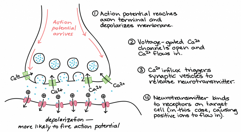
Communication at chemical synapses requires release of neurotransmitters. When the presynaptic membrane is depolarized, voltage-gated Ca2+ channels open and allow Ca2+ to enter the cell. The calcium entry causes synaptic vesicles to fuse with the membrane and release neurotransmitter molecules into the synaptic crevice. The neurotransmitter diffuses across the synaptic cleft and binds to ligand-gated ion channels in the postsynaptic membrane, resulting in a localized depolarization or hyperpolarization of the postsynaptic neuron. Image credit: Khan Academy https://world wide web.khanacademy.org/science/biology/ap-biological science/man-biology/neuron-nervous-system/a/the-synapse
- Excitatory postsynaptic potentials (EPSPs) make a postsynaptic neuronmore probable to burn down an action potential. For example, when acetylcholine is released at the synapse between a nerve and muscle (called the neuromuscular junction) past a presynaptic neuron, it causes postsynaptic Na+ channels to open up. Na+ enters the postsynaptic cell and causes the postsynaptic membrane to depolarize.
- Inhibitory postsynaptic potentials (IPSPs) make a postsynaptic neuronless likely to burn an activity potential. For example, when the neurotransmitter GABA (gamma-aminobutyric acrid) is released from a presynaptic neuron, it binds to and opens Cl– channels. Cl– ions enter the cell and hyperpolarizes the membrane.
Once neurotransmission has occurred, the neurotransmitter must be removed from the synaptic scissure then the postsynaptic membrane tin can "reset" and be ready to receive some other indicate. This can be accomplished in three ways:
- the neurotransmitter can diffuse abroad from the synaptic cleft
- the neurotransmitter can be degraded by enzymes in the synaptic cleft
- the neurotransmitter can be recycled (sometimes called reuptake) by the presynaptic neuron.
This video walks through the process of signal communication across a chemical synapse:
While activeness potentials are "all-or-nix," as noted above, EPSPs and IPSPs are graded; they vary in magnitude of depolarization or hyperpolarization, every bit illustrated below:
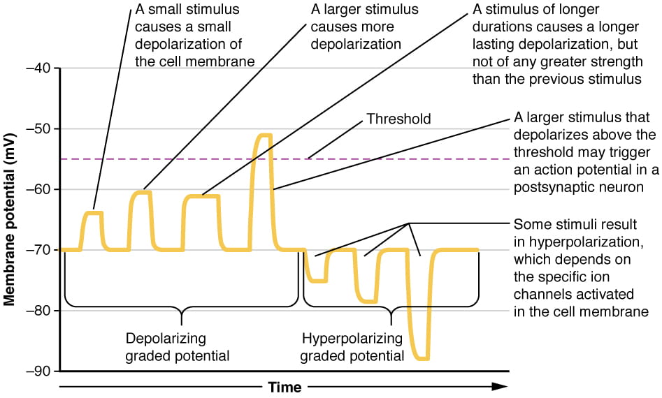
Graded potentials are temporary changes in the membrane voltage, the characteristics of which depend on the size of the stimulus. Some types of stimuli cause depolarization of the membrane, whereas others crusade hyperpolarization. It depends on the specific ion channels that are activated in the cell membrane. Image credit: OpenStax Anatomy & Physiology
Often a single EPSP is non strong enough to induce an action potential in the postsynaptic neuron on its own, and multiple presynaptic inputs must create EPSPs around the same time for the postsynaptic neuron to be sufficiently depolarized to fire an action potential. This process is called summation and occurs at the axon hillock, as illustrated below. In addition, each neuron oftentimes has inputs from many presynaptic neuron – some excitatory and some inhibitory – so IPSPs can cancel out EPSPs and vice versa. Information technology is the internet change in postsynaptic membrane voltage that determines whether the postsynaptic jail cell has reached its threshold of excitation needed to fire an action potential. Together, synaptic summation and the threshold for excitation human action as a filter so that random "noise" in the arrangement is non transmitted as important data.
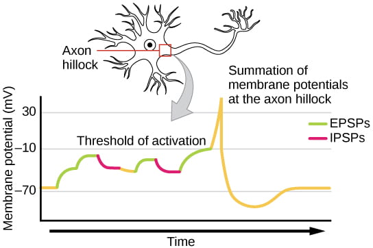
A unmarried neuron can receive both excitatory and inhibitory inputs from multiple neurons, resulting in local membrane depolarization (EPSP input) and hyperpolarization (IPSP input). All these inputs are added together at the axon hillock. If the EPSPs are strong enough to overcome the IPSPs and reach the threshold of excitation, the neuron will burn. Epitome credit: OpenStax Biology
This video, added afterwards the IKE was opened, provides an overview of summation in time and space:
Here are two final videos to help you put this all together (in a more engaging way than whatsoever of the videos higher up). Note that these videos practice non provide whatsoever new data, only they may help you lot ameliorate integrate all the information previously discussed:
kulikowskidins1974.blogspot.com
Source: https://organismalbio.biosci.gatech.edu/chemical-and-electrical-signals/neurons/
0 Response to "What Occurs to a Neuron When It Receives a Chemical Message"
Post a Comment